New Electrode System Offers a Window into Spinal Cord Function
FutureNeuro researchers, Dr Bahman Nasseroleslami and Dr Prabhav Mehra, at Trinity College Dublin have developed a new way to record spinal cord activity that could transform how scientists understand and track neurological conditions.
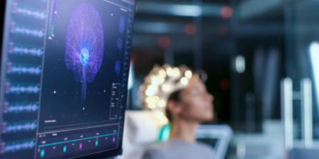
Their study, published in Clinical Neurophysiology, introduces a standardised, high-density electrode system (HD-ESG) that makes it possible to reliably and reproducibly measure electrical signals from the cervical spinal cord — the upper region of the spine that plays a crucial role in controlling movement.
A new way to understand the body’s motor networks
“We generally take our everyday motor functions for granted,” explained Dr Mehra, senior author of the study. “What feel like simple, basic movements are actually comprised of complex sensory-motor neuronal networks working in harmony across the brain, spinal cord, and peripheral nerves.”
One of the major challenges in studying spinal cord dysfunction is what researchers call the “clinico-radiological paradox.” In many neurological conditions, changes seen on spinal MRI scans — such as shrinkage or lesions — don’t always match the level of disability a person experiences, even when there’s strong clinical evidence of spinal involvement. HD-ESG offers a way to bridge this gap by measuring how the spinal cord actually functions, rather than just how it looks. By capturing detailed electrical activity in real time, it provides a window into the functional changes that underlie spinal cord damage and could one day offer new, quantitative markers to track disease progression.
In conditions like multiple sclerosis, these intricate circuits become disrupted, leading to progressive functional decline. The team uses electrophysiology techniques to characterise alterations in these multi-level sensory-motor communication pathways, mapping how signals are disrupted at different levels of the nervous system to uncover disease-related markers that can guide more precise treatments and personalised patient care.
Until now, disruptions at the spinal cord level have largely remained underexplored.
“Our recent publication on standardising the high-density recording of spinal cord signals is a key step toward addressing this gap and enabling reliable investigation of spinal cord dysfunction,” said Dr Mehra.
Developing a standardised system
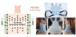
The team’s innovation—known as the SC10X/U electrode placement system—was motivated by a critical need for consistency in spinal cord research.
“A comparable and reproducible methodology is essential for research targeting clinical applications,” said Dr Mehra. “Currently, the lack of standardisation of spinal electrophysiology methodology has been a significant barrier to large-scale spinal neurophysiological studies. We believe that the SC10X/U electrode system will usher in a new wave of collaborative spinal research with greater translational potential.”
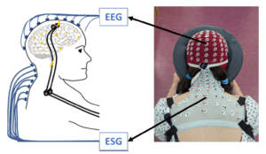
The new ESG electrode patch—the physical realisation of the SC10X/U system—works on a similar principle to how an EEG cap implements the 10-20 electrode system for brain recordings. Made from stretchy, breathable material in multiple sizes, it fits precisely along the neck and upper spine using key anatomical landmarks. Electrodes are filled with conductive gel for clear recordings and secured with fasteners and elastic bands.
Unlike previous methods, the patch can record up to 76 channels at once, giving much higher detail. Each channel is always placed in the same position, making it possible to compare data across different people and studies—something that wasn’t possible before.
Unlocking insights in multiple sclerosis
The HD-ESG system is already being used in MS studies at Trinity to find markers of spinal cord function that could help predict disease progression and guide treatment.
MS significantly affects the spinal cord, especially the neck region, and damage there is linked to worsening disability. But current imaging methods can’t fully track how the disease progresses over time. To address this, the team are using HD-ESG to measure spinal cord activity in response to stimulation. Early results suggest that these functional measurements may detect changes in the spinal cord more sensitively than traditional imaging, offering new ways to monitor MS and tailor treatments.
Broader clinical applications
Beyond MS, the researchers see significant potential for the technology across a range of neurological and neuromuscular conditions affecting the spinal cord, such as motor neuron disease, stroke, spinal cord injury and cerebral palsy.
He believes the field is entering a new era of discovery.
“The past couple of years have seen remarkable advances not only in neuroelectric spinal imaging like ours, but also in functional spinal MRI. I believe we are at the beginning of a new era of research focusing on spinal cord dysfunction and the neurophysiology of spinal circuits, and crucially, how they relate to wider sensory-motor networks. The implications of this will lead to more informed clinical care and rehabilitation strategies that move beyond just the cortex to a wider multi-system view.”
From prototype to global collaboration
The team’s next goal is to scale the SC10X/U electrode patch for large research studies and international collaboration.
“Our vision is to make the SC10X/U system widely accessible for any research group worldwide interested in advancing ESG research,” said Dr Mehra.
They are seeking industry partners to handle manufacturing, quality control, and global distribution, while the Trinity team provides technical support and scientific guidance. Interest is already growing, with multiple labs across Europe reaching out about using the patch.
Standardising ESG research has the potential to transform spinal cord studies in the same way EEG transformed brain research, enabling large-scale collaboration and more reliable results worldwide.